
Spectroscopically Well-Characterized RGD Optical Probe as a Prerequisite for Lifetime-Gated Tumor Imaging
15-Dec-2011
Molecular Imaging, 2011, DOI 10.2310/7290.2011.00018, Vol 10, No 6, pp 469–480 published on 15.12.2011
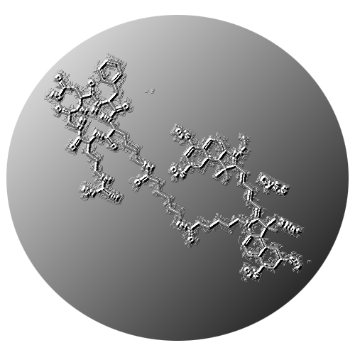
Labeling of RGD peptides with near-infrared fluorophores yields optical probes for noninvasive imaging of tumors overexpressing alpha-v-beta3 integrins. An important prerequisite for optimum detection sensitivity in vivo is strongly absorbing and highly emissive probes with a known fluorescence lifetime. The RGD-Cy5.5 optical probe was derived by coupling Cy5.5 to a cyclic arginine–glycine–aspartic acid–D-phenylalanine–lysine (RGDfK) peptide via an aminohexanoic acid spacer. Spectroscopic properties of the probe were studied in different matrices in comparison to Cy5.5. For in vivo imaging, human glioblastoma cells were subcutaneously implanted into nude mice, and in vivo fluorescence intensity and lifetime were measured. The fluorescence quantum yield and lifetime of Cy5.5 were found to be barely affected on RGD conjugation but dramatically changed in the presence of proteins. By time domain fluorescence imaging, we demonstrated specific binding of RGD-Cy5.5 to glioblastoma xenografts in nude mice. Discrimination of unspecific fluorescence by lifetime-gated analysis further enhanced the detection sensitivity of RGD-Cy5.5-derived signals. We characterized RGD-Cy5.5 as a strongly emissive and stable probe adequate for selective targeting of alpha-v-beta3 integrins. The specificity and thus the overall detection sensitivity in vivo were optimized with lifetime gating, based on the previous determination of the probes fluorescence lifetime under application-relevant conditions.